Electrocardiography is a medical technique by which minute electrical impulses, related to the functioning of the heart muscle, are detected, recorded, and printed out over time for interpretation by doctors. It is the best way to detect, diagnose, and determine the severity of many heart problems, mostly involving abnormal heart rhythms. It is also useful for detecting certain types of heart damage, murmurs, and circulatory problems.
The term electrocardiography is derived from three Greek words relating to electricity, the heart, and writing. The printout of data obtained from electrocardiography is called an electrocardiogram and is often abbreviated as ECG or EKG. Many doctors and health professionals prefer EKG as it helps prevent confusion with another type of medical process called an electroencephalogram or EEG.
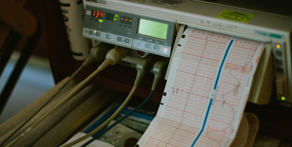
A patient being examined by electrocardiography is fitted with a number of skin electrodes, sensors which can detect the minute electrical impulses of the human body. These electrodes are placed at various points on the body, mostly on the chest, but also at each wrist and ankle. They transmit the electrical impulses generated by the operation of the heart and circulatory system back to a central unit that interprets the data and prints it out in a continuous, real-time format on paper. Many machines also have a digital display and recording capability.
A doctor can study the data on an EKG and use it to diagnose and detect irregular heart rhythms, certain types of heart damage, and other circulatory problems. An EKG is especially useful for diagnosing damage due to myocardial infarctions, commonly known as heart attacks. Patients believed to have suffered a heart attack or in danger of an imminent heart attack are nearly always hooked up to an electrocardiography machine as soon as they come under medical care.
Doctors rely on electrocardiography to give them much information that would be unavailable without surgery or more invasive procedures. A skilled cardiologist, or doctor that specializes in treating heart problems, can obtain a great deal of information about a patient's heart and circulatory system through electrocardiography. Even certain genetic abnormalities or the presence of some types of drugs may be detectable through EKG analysis.
An EKG is often used as a monitoring tool for patients with heart or circulatory problems, in addition to its use as a diagnostic tool. It is not uncommon for these types of patients to be connected to an EKG machine for extended periods while under care or treatment for these types of issues. Alarms may be programmed to alert doctors to potential problems that may arise with these patients while connected to one of these machines.
Types of electrocardiograms
Stress test
Some heart problems only appear during exercise. During stress testing, you’ll have a continuous EKG while you’re exercising. Typically, this test is done while you’re on a treadmill or stationary bicycle.
Holter monitor
Also known as an ambulatory ECG or EKG monitor, a Holter monitor records your heart’s activity over 24 to 48 hours or up to 2 weeks while you maintain a diary of your activity to help your doctor identify the cause of your symptoms. Electrodes attached to your chest record information on a portable, battery-operated monitor that you can carry in your pocket, on your belt, or on a shoulder strap.
Event recorder
Symptoms that don’t happen very often may require an event recorder. It’s like a Holter monitor, but it records your heart’s electrical activity just when symptoms occur. Some event recorders activate automatically when they detect arrhythmia. Other event recorders require you to push a button when you feel symptoms. You can send the information directly to your doctor over a phone line.
Loop recorder
A loop recorder is a device that is implanted in your body, under the skin of your chest. It functions as an electrocardiogram would but allows for continuous remote monitoring of your heart’s electrical signals. It’s looking for irregularities that can cause fainting or palpitations.
Why it's done?
An electrocardiogram is a painless, noninvasive way to help diagnose many common heart problems. A health care provider might use an electrocardiogram to determine or detect:
- Irregular heart rhythms (arrhythmias)
- If blocked or narrowed arteries in the heart (coronary artery disease) are causing chest pain or a heart attack
- Whether you have had a previous heart attack
- How well certain heart disease treatments, such as a pacemaker, are working
You may need an ECG if you have any of the following signs and symptoms:
- Chest pain
- Dizziness, lightheadedness or confusion
- Heart palpitations
- Rapid pulse
- Shortness of breath
- Weakness, fatigue or a decline in ability to exercise
What can you expect?
Before
You may be asked to change into a hospital gown. If you have hair on the parts of your body where the electrodes will be placed, the care provider may shave the hair so that the patches stick.
Once you're ready, you'll typically be asked to lie on an examining table or bed.
During
During an ECG, up to 12 sensors (electrodes) are attached to the chest and limbs. The electrodes are sticky patches with wires that connect to a monitor. They record the electrical signals that make the heartbeat. A computer records the information and displays it as waves on a monitor or on paper.
You can breathe during the test, but you will need to lie still. Make sure you're warm and ready to lie still. Moving, talking or shivering may interfere with the test results. A standard ECG takes a few minutes.
After
You can typically return to your usual activities after your electrocardiogram
Results
ECG results can give a health care provider detail about the following:
- Heart rate. Usually, heart rate can be measured by checking the pulse. An ECG may be helpful if your pulse is difficult to feel or too fast or too irregular to count accurately. An ECG can help identify an unusually fast heart rate (tachycardia) or an unusually slow heart rate (bradycardia).
- Heart rhythm. An ECG can detect irregular heartbeats (arrhythmias). An arrhythmia may occur when any part of the heart's electrical system doesn't work properly.
- Heart attack. An ECG can show evidence of a previous heart attack or one that's currently happening. The patterns on the ECG may help determine which part of the heart has been damaged, as well as the extent of the damage.
- Blood and oxygen supply to the heart. An ECG done while you're having symptoms can help your health care provider determine whether reduced blood flow to the heart muscle is causing the chest pain.
- Heart structure changes. An ECG can provide clues about an enlarged heart, heart defects and other heart problems.
What risks are involved?
There are few, if any, risks related to an EKG. Some people may experience a skin rash where electrodes were placed, but this usually goes away without treatment.
People undergoing a stress test may be at risk for a heart attack, but this is related to the exercise, not the EKG.
An EKG only monitors the electrical activity of your heart. It does not emit any electricity and is completely safe, even during pregnancy.
The Holter monitors can sometimes cause an allergy or rash on the areas of the skin where the EKG electrode pads are placed. This is more likely when they’re worn for many consecutive days.
Loop recorders are often used without any negative effects and have gotten smaller and more efficient over time. As with any procedure like this one, there is the possibility of mild pain, slight bruising, or infection at the implantation location.


