Back when medical technology was not as developed as it is today, physicians had to ‘feel’ the affected organ of their patients’ bodies and then use their experience to determine the underlying cause of the discomfort.
Having no other form of examination at their disposal, our ancient ancestors had to depend entirely on this crude, rudimentary technique of detecting problems inside the body. Fast forward to today, and we can spot the smallest malignant growth anywhere inside the human body, thanks to ultrasound imaging.
What is Ultrasound Scanning?
Ultrasound scanning, or simply ultrasound, is a non-invasive and painless diagnostic technique used to “look inside” the innards of a human body. Also known as sonography, it’s quite an effective and widely used technique that doctors and medical experts use to determine what’s happening inside the human body without actually operating on it. Although it can be (and is) used in nearly all cases where the state of a patient’s internal organs must be determined (such as to check blood circulation in a limb, detection of tumors etc.), sonography is most often associated with pregnancy, as it helps to determine various parameters of fetal development, which helps to calculate the due date.
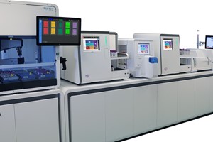
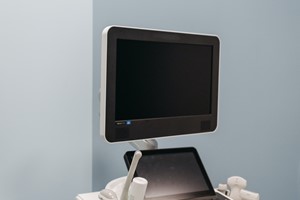
The Ultrasound machines
Unlike an X-ray machine, which uses electromagnetic waves to form images of the human body, ultrasound machines use sound; more specifically, it uses ultrasound waves (hence the name ‘ultrasound machine’) to find out what’s going on inside the body.
Structurally, an ultrasound machine is very simple. It consists of two basic parts:
- A transducer probe that sends sound waves and receives their echoes
- A computer (a central processing unit or CPU) that supplies power to the transducer probe, performs all the calculations and shows the images of the internal area on a screen.
How does sonography work?
The transducer probe comes in various shapes and sizes. Irrespective of their appearance, most of them contain one or more quartz crystals called piezoelectric crystals. The interesting thing about these crystals is that they change shape, or vibrate, when an electric current is applied to them. So, when the transducer is switched on, the crystals create vibrations that produce sound waves with frequencies (anywhere between 1 and 18 MHz) that are higher than what humans can hear.
Before starting the process, a water-based gel is applied directly to the skin on the designated area of the subject’s body. This avoids the formation of extremely tiny air bubbles between the probe and the skin, which could potentially hamper the transmission and reception of sound waves. The sound beam produced from the probe is focused either by the shape of the probe itself, a lens in front of the probe, or a set of control pulses from the scanner, resulting in an arc-shaped sound wave emanating from the face of the probe, entering the body and finally coming into focus at the desired depth.
The transmitted sound waves pass through the thin layer of skin, but bounce off fluids, tissues and internal organs. These reflected waves are received by the probe, which converts them into electric signals (thank you. piezoelectric crystals!) and feeds them into the computer. The computer uses this information and performs quick calculations (a lot of them!) using the speed of sound and the time taken by the echo to reach the probe to create a real-time image of the inside of the human body.
Traditional ultrasound scanners display 2-dimensional images, meaning that images are displayed in flat sections of the body. However, modern scanners use the same calculations to produce 3-dimensional images, which is a wonderful advancement, especially for would-be parents watching the growth of their baby.
There are a few different types of ultrasonography, depending on the type of examination required, including Doppler ultrasonography (to determine whether certain structures are moving away from or towards the probe), contrast ultrasonography (used in echocardiography and for lesion characterization), and molecular ultrasonography.
If not for this simple, yet incredibly effective machine, not only would we be unable to locate a tumor or diagnose a disease inside the human body, but we would be deprived of the heartwarming sight of watching a new life grow inside the womb.
The global ultrasound device market size is expected to reach USD 13.56 billion by 2028, according to a new report by Grand View Research, Inc. The market is expected to expand at a CAGR of 4.8% from 2021 to 2028. Ultrasound is a very efficient technology that aids in the diagnosis of various disorders such as tumors as well as the detection of changes in the appearance of organs, tissues, and arteries. It also has a range of therapeutic applications. An increase in the adoption of ultrasound devices for diagnosis and treatment is driving the market. In addition, a rise in the adoption of minimally invasive surgeries, coupled with technological advancements, is expected to boost market growth.
Ultrasound Device Market Growth & Trends
Emerging innovations in ultrasound technology are expanding the market size. Hand-held ultrasound devices have made the technology more accessible for use in limited-resource communities around the world. Furthermore, the introduction of 3D/4D ultrasound and the integration of artificial intelligence (Al) to automate time-consuming processes are expected to propel market growth during the forecast period. For instance, in September 2020, GE Healthcare launched Voluson SWIFT, a new ultrasound technology, which is designed to help women's health by allowing clinicians to enhance patient outcomes by expanding diagnostic capabilities. The system includes industry-first AI algorithms for auto-recognition and has excellent image quality.
The rising prevalence of chronic diseases like cancer, neurological conditions, orthopedic conditions, and cardiovascular diseases, which demand the adoption of ultrasound devices for diagnosis, is expected to expand the market during the forecast period. In addition, the market for therapeutic ultrasound equipment is predicted to grow due to the rising use of ultrasound techniques for various therapeutic procedures using High-Intensity Focused Ultrasound (HIFU) technology and Extracorporeal Shockwave Lithotripsy (ESWL).