In a recent study published in Biomedicine and Pharmacotherapy, researchers have outlined a groundbreaking therapeutic approach for neonatal hypoxic-ischemic (HI) brain injury, offering promising advancements in the field of pediatric neurology.
Background:
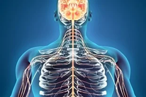
Neonatal hypoxic-ischemic encephalopathy (HIE) arises due to the deprivation of oxygen and blood flow to the brain during labor and delivery. It stands as a significant contributor to cerebral palsy, mortality, and other neurological disorders among infants.
Therapeutic hypothermia (TH) has long been the standard of care for HIE. However, TH presents several limitations and challenges in clinical application, including cognitive impairments, variable clinical responses, and incomplete protection, necessitating the development of more effective and accessible therapies for HIE.
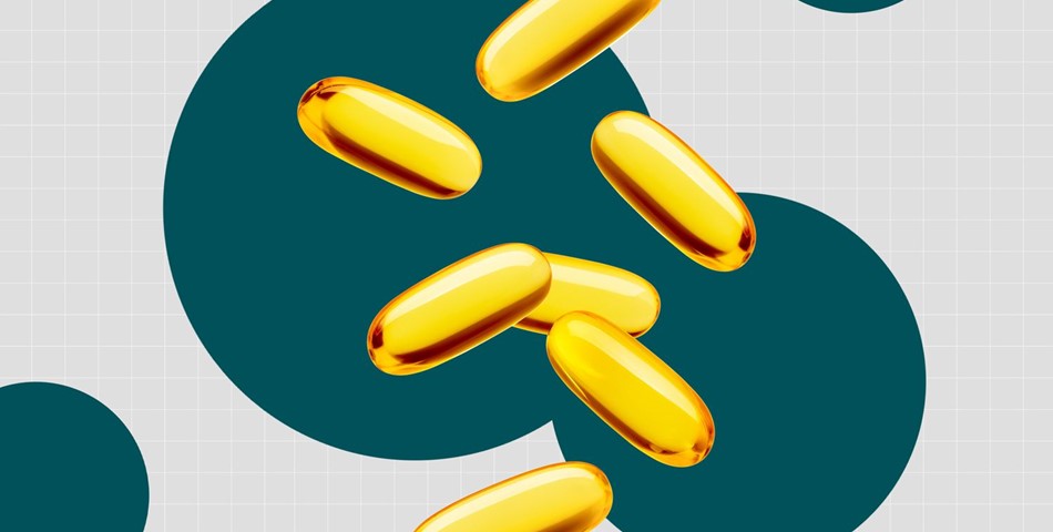
Studies have suggested that dietary supplements and oral omega-3 fatty acids (n-3 FAs) hold promise as potential neuro- and cardioprotective strategies.
Previously, researchers demonstrated that administering docosahexaenoic acid (DHA) as triglyceride (TG) emulsion particles following HI injury in neonatal mice significantly mitigated brain damage.
About the Study:
In the present study, researchers investigated the efficacy of a novel n-3 diglyceride (DG) lipid emulsion in neonatal HI brain injury. They produced n-3 DG oils through reverse glycerolysis reactions and prepared 10% lipid emulsions by mixing n-3 DG or TG-DHA oils with egg yolk phosphatidylcholine.
The average particle size, polydispersity index (PDI), and zeta potential of the emulsions were determined. Oxidative measurements were conducted using the p-anisidine assay.
The emulsions were incubated with a buffer with or without bovine lipoprotein lipase (LpL). Human plasma containing apolipoprotein C-II was introduced into the mixture, and released free fatty acid (FFA) was measured.
Experiments were performed with increasing LpL levels. C57BL/6J neonatal mice (eight days old) were intraperitoneally injected with DG or TG emulsion, and changes in plasma glyceride levels were evaluated. Blood samples were collected after injection.
Furthermore, another group of mice aged 10 days underwent HI insult. Mice received two doses of DG or TG emulsion, one shortly after the HI insult and the other an hour later; controls were injected with saline (two doses).
Moreover, Wistar rat pups aged seven days also underwent a similar procedure for HI brain injury. Rats received a single dose of saline, n-3 DG, or Omegaven (commercial TG-based emulsion) immediately after the HI insult.
One group of rats received TH for five hours post-HI insult. Negative geotaxis and righting reflex performances were evaluated 24 hours after the injury. Twenty-four hours after reperfusion, mouse brains were obtained (immediately after behavioral tests), and infarct sizes were computed.
Eight days after reperfusion, rat brains were obtained; hematoxylin and eosin staining were performed to analyze brain area loss. Immunofluorescence was conducted for astrocyte and microglia markers.
Findings:
n-3 DG preparations exhibited smaller particle sizes than Omegaven or n-3 TG. Both n-3 DG and n-3 TG emulsions were homogeneous with low PDI values. Moreover, n-3 TG emulsions displayed FFAs at 4–8 weeks, indicating spontaneous hydrolysis, while n-3 DG emulsions did not demonstrate such deterioration.
Additionally, there was no detectable deterioration in the DG emulsion at six months. The p-anisidine values of all oils and emulsions were below 20 mEg/l.
The zeta potential was -50 mV for DGs and -35 mV for TGs. Basal FFA levels (without LpL) were similar between DG and TG emulsions. The highest lipolysis was observed in the n-3 DG emulsion, with over 1.5-fold more FFAs released than in n-3 TG emulsions.
Plasma glyceride levels in neonatal mice were substantially elevated one hour after n-3 DG injection, peaking at two hours with a three-fold increase compared to baseline, and returning to baseline levels by four hours.
In contrast, glyceride levels at one hour after n-3 TG injection were similar to baseline levels but increased at later time points (2h and 4h).
Moreover, neonatal HI injury mice treated with n-3 DG exhibited a significant decline in infarct size (87%), whereas n-3 TG treatment reduced the damage by 43%. Similarly, n-3 DG proved to be the most effective in the rat model.
Furthermore, the reflex performance of neonatal HI mice after n-3 DG treatment was akin to age-matched naïve mice, suggesting that n-3 DG preserved neurofunctional outcomes.
In the rat model, astrogliosis significantly reduced seven days after HI injury with n-3 DG treatment compared to saline. Moreover, microgliosis was also significantly attenuated in the n-3 DG treatment group relative to the saline group.
Conclusions:
The research has indicated that n-3 FAs in DG lipid emulsions offer greater benefits than n-3 TG in reducing brain injury.
The n-3 DG emulsion outperformed TH, the current standard of care, in decreasing infarcts, and it also attenuated astrogliosis and microgliosis during the sub-acute phase of the injury. Therefore, n-3 DG confers neuroprotection and activates cytoprotective mechanisms in response to brain injury.
By: Tarun Sai Lomte
https://www.news-medical.net/